Key points
- Source localization estimates where the activity measured at the surface of the head arises. The problem has no unique solution: each method adds its own assumptions.
- Three families: dipoles, distributed methods (minimum norm, LORETA, sLORETA) and beamformers.
- Three conditions weigh as much as the method: the number and coverage of electrodes, the actual sensor positions and the head model.
- With 64 to 256 electrodes, precision becomes sublobar, according to the International Federation of Clinical Neurophysiology.
The inverse problem
Computing surface potentials from a known source in the brain is the forward problem: it has a solution. The reverse path has no unique one. The same potential map can result from an infinite number of source configurations.
To choose among them, a constraint has to be added: assume that there are only a few sources, or that activity is as weak as possible, or that it varies smoothly from one point to its neighbours. Each method corresponds to one of these constraints. The result is an estimate, whose quality depends on how valid the assumption is for your data.
The six steps
A practical review (Michel and Brunet, 2019) describes the chain that leads from the recording to a three-dimensional image of neuronal activity.
- A clean signal: artefacts removed, bad channels interpolated, average reference.
- Electrode positions: measured on each participant, or taken from a template.
- The head model: the geometry and conductivity of the brain, skull and scalp.
- The source space: the points in the brain where activity is allowed, generally the grey matter.
- The inverse method: the computation that distributes activity among those points.
- Analysis: regions of interest, statistics, connectivity between sources.
From the recording to the source map
- 1Clean signalArtefacts removed, average reference.
- 2Electrode positionsMeasured or taken from a template.
- 3Head modelGeometry and conductivity.
- 4Source spaceThe points where activity is allowed.
- 5Inverse methodThe computation that distributes activity.
- 6AnalysisRegions, statistics, connectivity.
Families of methods
| Method | Assumption | Suited to | Limit |
|---|---|---|---|
| Equivalent dipole | Activity comes from a small number of point sources | Single focal source: epileptic spike, early sensory response | The number of dipoles has to be fixed; unsuited to extended activity |
| Minimum norm (MNE) | Among the possible solutions, the one with the lowest energy is chosen | Distributed activity, with no assumption about the number of sources | Favours superficial sources |
| LORETA | Activity varies smoothly from one point to its neighbours | Extended activity | Blurred images, low spatial resolution |
| sLORETA, eLORETA | Standardised current density | Locating the activity maximum | Images are still smeared; the stated precision holds for a point source |
| Beamformer | Sources are not correlated with one another | Oscillatory activity, connectivity | Underestimates strongly synchronised sources |
The author of sLORETA presents it as a linear solution whose localization error is zero for a point source. That is a mathematical property in an ideal, noise-free setting; it does not remove the need for the other conditions.
How many electrodes?
This is the question on which the data are clearest.
- Lantz et al., 2003. In 14 epileptic patients recorded with 123 electrodes, the authors repeated the localization with 63 and then 31 electrodes. The source was significantly closer to the lesion with 63 than with 31 electrodes in 9 patients, and in 11 with 123 electrodes.
- Song et al., 2015. Simulations and epilepsy data show that higher sensor density improves precision, and that adding sensors on the lower part of the head improves the estimate at all depths. The authors work at EGI.
- Seeck et al., 2017. The International Federation of Clinical Neurophysiology writes that arrays of 64 to 256 electrodes allow source imaging with sublobar precision. It adds that young children need at least as many electrodes as adults.
Two ideas to remember: density matters, and so does coverage. Sensors on the face, the lower temples and the neck inform about deep and basal sources, which the top of the head alone sees poorly.
The head model
Current passes through the brain, the fluid, the skull and the scalp before reaching the electrodes. The skull, a poor conductor, smears the potentials. The head model describes this path.
- Concentric spheres: simple, fast, not very faithful to anatomy.
- Boundary elements: the surfaces of the brain, skull and scalp, taken from an MRI.
- Finite elements or finite differences: a volume mesh that accounts for variations in thickness and conductivity.
Each participant's own MRI gives the best model. Failing that, a template head fitted to the actual electrode positions remains usable. In every case, knowing the position of each sensor on the participant's head reduces an avoidable source of error.
Limits to keep in mind
- It is not functional MRI. The images are smeared. Localization points to a region, not a point.
- Deep sources are estimated less well than superficial cortical sources.
- Skull conductivity varies from person to person and with age. It is rarely measured.
- Noise propagates: a poorly removed artefact becomes a false source.
- The choice of method steers the result. Presenting two methods, or comparing with another modality, strengthens a conclusion.
Software tools
Michel and Brunet's review illustrates each step with Cartool, a free academic software package. Other open tools are widely used in research: Brainstorm, FieldTrip, MNE-Python and EEGLAB. Manufacturers also offer their own modules.
Before choosing, check three points: is your file format read directly, is your sensor layout provided, and can you import measured positions and your participants' MRI.
With an EGI system
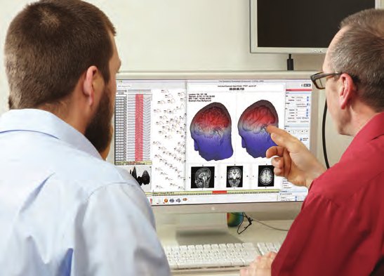
- Up to 256 channels, with sensors evenly distributed over the whole head, including the face and neck.
- 128 channels from infancy, in line with the recommendation not to reduce the number of electrodes in children.
- Sensor digitisation: the manufacturer offers a photogrammetry system to measure the position of each sensor.
- Tools: the manufacturer has a source estimation module; Net Station files export to MATLAB, EEGLAB and FieldTrip.
- In France: the LTSI team in Rennes published in 2014 a source connectivity method based on high-resolution EGI EEG recordings.
Frequently asked questions
What is the best source localization method?
There is no single best one. A dipole suits a single focal source, such as an epileptic spike or an early sensory response. Distributed methods, minimum norm, LORETA or sLORETA, suit extended or multiple activity. Beamformers suit oscillatory activity. Many teams compare two methods.
How many electrodes are needed to localize sources?
The more there are, the more precise the localization. In a study of 14 epileptic patients recorded with 123 electrodes, the source was closer to the lesion with 63 electrodes than with 31 in 9 patients, and in 11 with 123 electrodes. The International Federation of Clinical Neurophysiology states that arrays of 64 to 256 electrodes allow source imaging with sublobar precision.
Is each participant's MRI needed?
It improves the head model, and therefore the localization. Failing that, a template head is used, fitted to the actual electrode positions. The choice depends on the precision sought and on access to MRI.
Are more electrodes needed in children?
At least as many as in adults, according to the International Federation of Clinical Neurophysiology: a child's skull is thinner, which makes potentials more focal at the surface and increases the risk of spatial undersampling.
Sources
The scientific publications come from PubMed.
- Michel CM, Brunet D. EEG Source Imaging: A Practical Review of the Analysis Steps. Front Neurol. 2019;10:325. doi:10.3389/fneur.2019.00325
- Lantz G, Grave de Peralta R, Spinelli L, Seeck M, Michel CM. Epileptic source localization with high density EEG: how many electrodes are needed? Clin Neurophysiol. 2003;114(1):63-9. doi:10.1016/s1388-2457(02)00337-1
- Song J, Davey C, Poulsen C, et al. EEG source localization: Sensor density and head surface coverage. J Neurosci Methods. 2015;256:9-21. doi:10.1016/j.jneumeth.2015.08.015
- Seeck M, Koessler L, Bast T, et al. The standardized EEG electrode array of the IFCN. Clin Neurophysiol. 2017;128(10):2070-2077. doi:10.1016/j.clinph.2017.06.254
- Pascual-Marqui RD. Standardized low-resolution brain electromagnetic tomography (sLORETA): technical details. Methods Find Exp Clin Pharmacol. 2002;24 Suppl D:5-12. PubMed 12575463
- Hassan M, Dufor O, Merlet I, Berrou C, Wendling F. EEG source connectivity analysis: from dense array recordings to brain networks. PLoS One. 2014;9(8):e105041. doi:10.1371/journal.pone.0105041
- Magstim EGI. “GES 400 MR” brochure, March 2017 (related products: GeoSource, Geodesic Photogrammetry System).
This article is general information for research teams. It is based on the manufacturers' documents and the abstracts of the publications cited. Gaze Intelligence distributes Magstim EGI systems: this article is written by a distributor.